xr lumbar spine ap and lateral
 Radiograph of the lumbar spine AP (A) and Lateral View (B) of 51 year... | Download Scientific Diagram
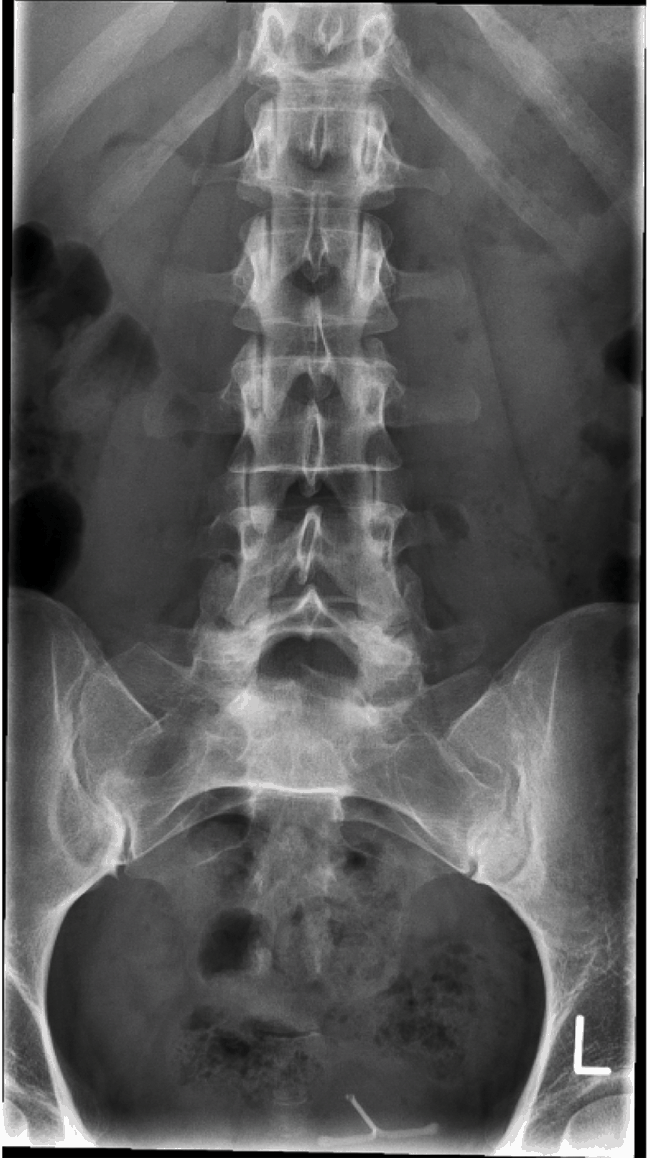
Username Password Remind me Username Password Remind me X-ray Posting of the lumbar columnBy: CE4RT Radiologists consider a good-quality spinal radiographic film of lumbar spine when it shows the lower ribs, the lumbar vertebral bodies, the transverse processes, the pédic, the spiny processes, the sacrolynic joints and the sacroiliac joints. In the lateral projections, the intervertebral spaces of the disc and intervertebral foramen, as well as the upper and lower joint processes, should be visible along with the vertebral bodies and the spiny processes. Lumbar Spine AP or PAPurpose and Structures Shown A basic view of the lumbar column. Patient position Supine or prone. The injured patients should not be returned. In patients with trauma, a lumbar X-ray is performed in the AP or PA position with a minimal movement of the patient. Part Position The patient's knees are bent to ensure that the back is flat on the table. The patient should be asked to stop breathing when the exposure is taken. Lumbar Spine PA Method FergusonPurpose and Structures Shown An alternative view of the lumbar column in the projection of the PA to protect the radiosensible organs from the exhibition. This is a series of scoliosis that is used to distinguish a primary deformation curve from a compensatory curve. Position of the patient Standing or sitting in front of a vertical grid. The RI must be adjusted to include about 1 inch of the illiterate ridges. The midsagittal plane of the body must be aligned to the median line of the network. The arms hung next to the body in the standing position. The elbows are flexed and the hands rest on the lap in the sitting position. A second X-ray is taken with the hip or the elevated foot with a block or sandbag below the foot or the buttock. The Ferguson method requires the patient to move some effort to maintain this position without support. Part Position The patient should be asked to suspend breathing during the exposure. Candy is protected. The image shows the chest and lumbar vertebrae in the PA projection with the spinal column in the center of the X-ray. Lumbar Spine LateralPurpose and Structures Shown A basic view of the lumbar column. Position of the patient lying on the left or right side (side mirror) with the knees and hips flexed for comfort. The elbows are flexed and the arms are at a straight angle to the body. The knees are superimposed. The midcoronal plane is aligned with the median network line. In the patient suspected of fracture, the spine lumbar with horizontal beam must be used instead. Position of the part The candies are protected. The patient should be asked to inspire, breathe and then hold the breath during the exposure. The image shows the lower area of the thoracic to the coccyx with lumbar vertebral bodies, intervertebral disk spaces, spiny processes and the visualized lumbosacral union. Lumbar Spine Lateral SupinePurpose and Structures Shown Additional view of the lumbar spine for patients with injuries. Position of the Supine patient with a horizontal beam. The midsagittal plane is centered in the center of the network. The hips and shoulders are on the same plane horizontally. The elbows are flexed and the hands are placed in the upper chest to remove the forearms from the field of exposure. The patient may be asked to bend the knees and hips to bring the back in firm contact with the table and reduce the lumbar lordosis. Position of the part The candies are protected. The patient should be asked to breathe and keep breathing during the exposure. The image shows the lumbar vertebral bodies, disc intervertebral spaces, cross-sectional and spiny processes, pedics and laminas. The sacroiliac joints are seen on both sides at the same distance from the spine. Vertebras are symmetrical with spiny processes focused on the bodies. In trauma patients, a larger field can be used to allow the visualization of additional organs such as the kidneys, liver, spleen, and muscle of the psoas. Lumbar Spine AP ObliquePurpose and Structures Shown An oblique projection that is usually made after the AP projection to demonstrate joint processes and/or lumbosacral processes. Patient position Supine and turn 45 degrees to the affected side. The long axis of the body must be parallel to the long axis of the table. The column is centered on the midline of the grid. The lumbar column is approximately 2 inches medium to the upper iliac column elevated in the oblique position. The arms are in a comfortable position. The patient should be asked to keep the breath during the exposure. Position of the part A support can be placed under the elevated parts of the body (shoulder, hip, knee). Candy is protected. The degree of rotation of the body must be 45 degrees to demonstrate the joint processes and 30 degrees to demonstrate the lumbosacral processes. The image shows an oblique projection of the area from the lower chest vertebrae to the sacrum. The joint processes are displayed on the closest side to the RI. Zygapophyseal joints are open and evenly seen through the vertebral bodies. The other side is visualized for comparison. Sacrum APPurpose and Structures Shown A basic view of the lumbosacral union and the sacroiliacal joints. Patient position Supine with a vertical beam at 15 degrees. This view is not used in children. Gas and fecal matter in the intestine can interfere with images of the sacrum and coccyx. Intestinal preparation may be required (by doctor's order). The bladder must be emptied before the test. Part Position The tube must be sharpened to 15 degrees. The center must be made 3 cm on pubic sympathies. The patient can breathe normally during exposure. Candy is protected in men. Female hens cannot be protected for this projection. The image clearly shows the sacrum free of superimposition. Lumbosacral Junction LateralPurpose and Structures Shown A basic view of the lumbosacral union. This view is not used in children. Position of patient lying on the left or right side. The patient should be asked to double the knees to stabilize the body. A pad must be placed under the waist for the support. If possible, the hips must be complete. Position of the part The centralization must be made 3 cm below the iliac crest. The knees are exactly overlapping. The patient should be asked to suspend breathing during the exposure. Candy is protected. The image shows the lumbosacral articulation in the center of the image. The entire L5 and the upper sacrum should be visualized. Hey, X-Ray Techs! The first time visitors get 1 CE Credit free. The first time visitors get 1 CE Credit free.1 Category A Credit to meet ARRT* requirements.$11.95 FREE! Our courses About X-Ray CE Credits Useful stuff Back to TopThank you for choosing CE4RT.com. Do you have any questions? Maybe you have suggestions on how to make our courses or site even better? We would love to hear from you, just give us a text or call to 657-222-0777 Copyright © 2021 XRC LLC All rights reserved. 848 N. Rainbow Blvd. #4152 - Las Vegas, NV 89107 657-222-0111* ARRT © is a registered trademark property of The American Registry of Radiologic Technologists. Although our courses are accepted by the ARRT for credit, as with all other EC providers online this website is not directly authorized by, supported by, or affiliated with the American Register of Radiological Technicians.

Lumbar spine (AP/PA view) | Radiology Reference Article | Radiopaedia.org
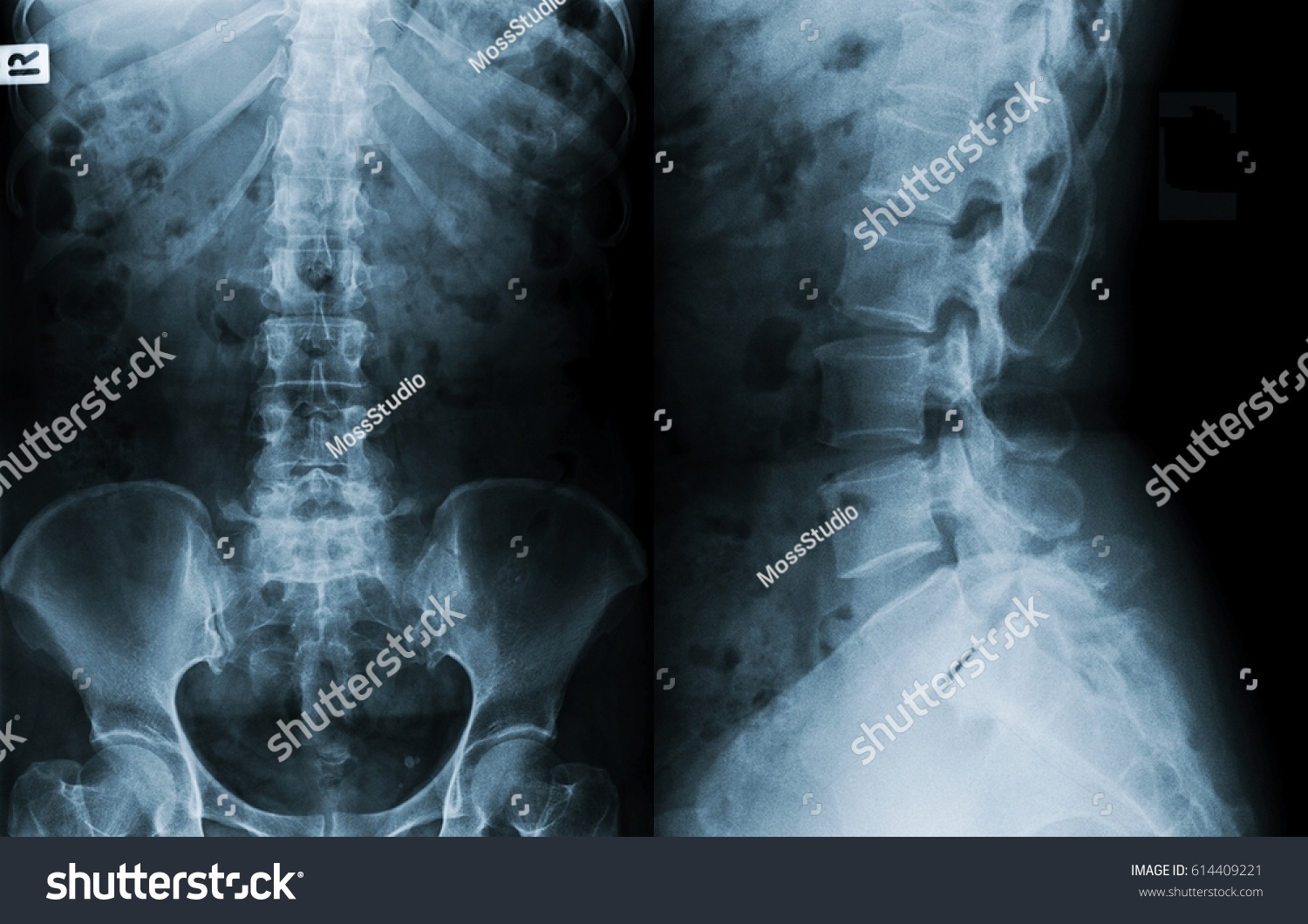
Film Xray Lumbar Spine Ls Spine Stock Photo (Edit Now) 614409221
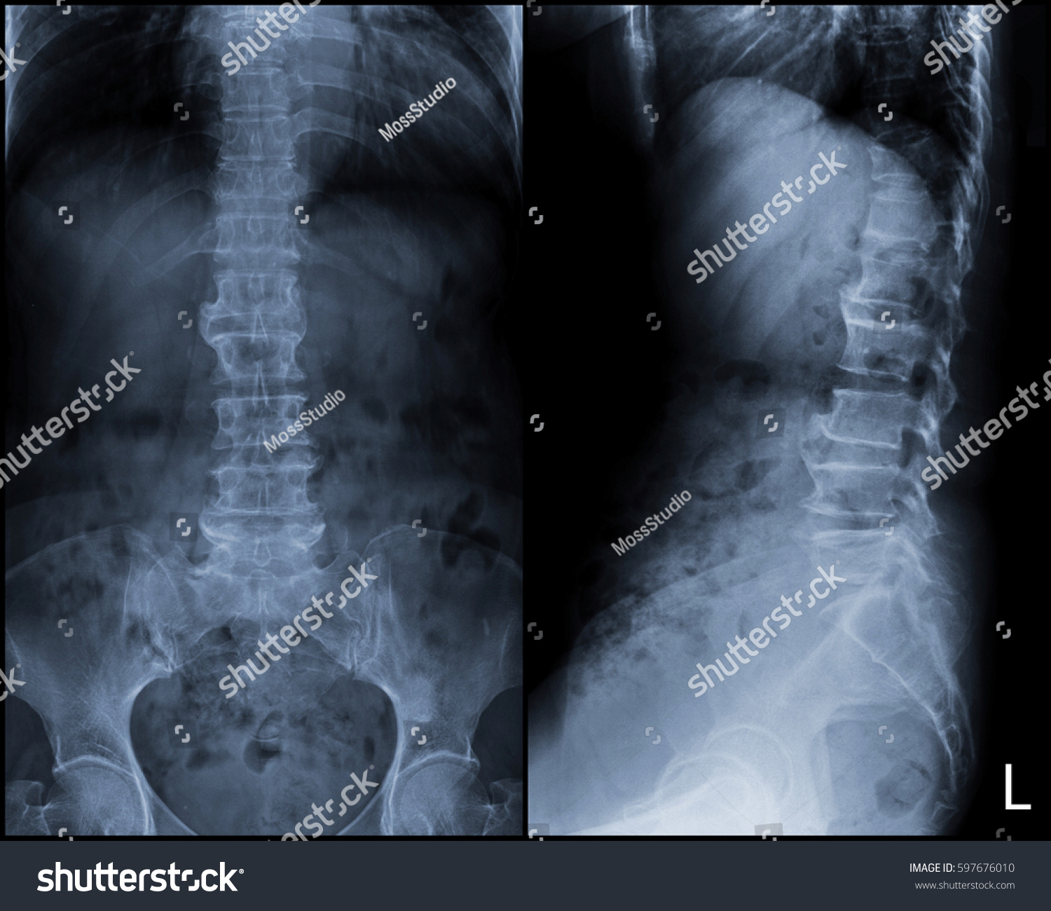
Film Xray Ls Spine Lumbar Spine Stock Photo (Edit Now) 597676010

Lumbar Spine Radiographic Anatomy | Radiology student, Diagnostic imaging, Medical knowledge

X-ray of lumbar spine antero-posterior (AP) and lateral views showing... | Download Scientific Diagram
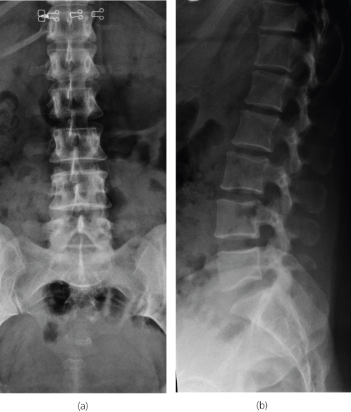
Thoracic and Lumbar Spine | Radiology Key
CE4RT - Radiographic Positioning of the Lumbar Spine for X-ray Techs
Shokeen X-ray & Dignostics Centre
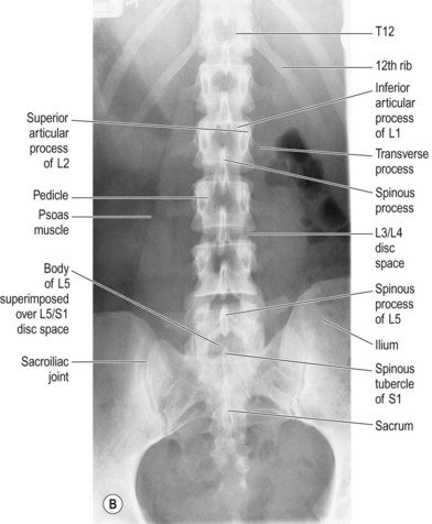
Lumbar spine | Radiology Key

Lumbar Spine-AP, Lateral and Oblique - YouTube
Lateral Lumbar Spine Radiography - wikiRadiography

Lumbar X-rays: A systematic approach | Neurosurgery Basics

X-ray L-S Spine AP,LATERAL: Finding Moderate Compression Fracture.. Stock Photo, Picture And Royalty Free Image. Image 133049474.

View Image
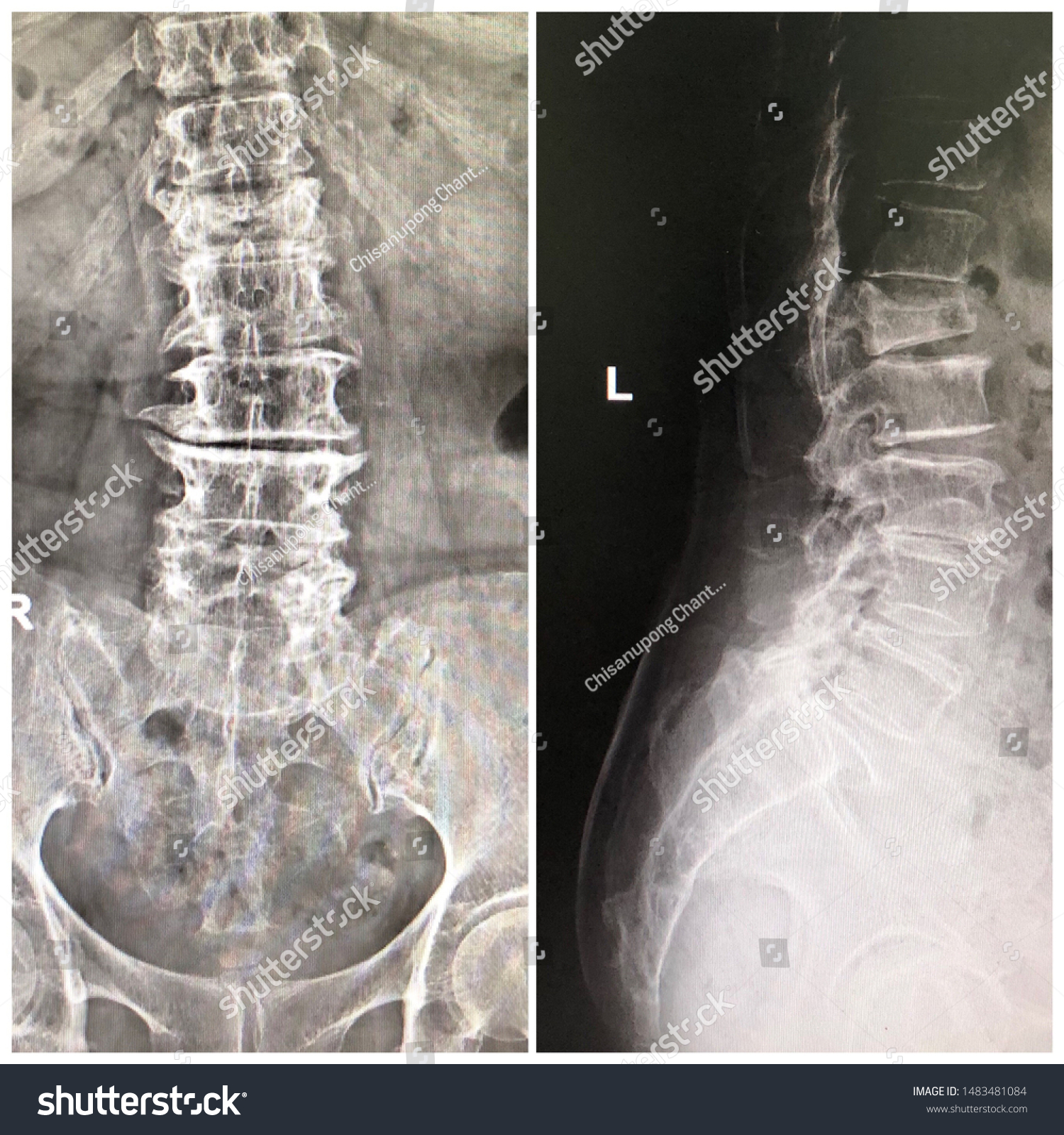
X Ray Lumbar Spine Ap Lateral Stock Photo (Edit Now) 1483481084

X-ray films of the lumbar spine. A) Anteroposterior (A-P) view. Slight... | Download Scientific Diagram
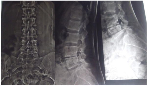
Spinal Tuberculosis – Directly Observed Treatment and Short Course or Daily Anti Tubercular Therapy -Are We Over Treating?

Radiographic Anatomy of the Skeleton: Lumbar Spine -- AP View, Labelled | Radiology humor, Radiology technician, Radiology schools
Lumbar Spine Radiography - wikiRadiography

LS Spine (AP/Lat) - Wellcare Diagnostic Clinic, Thane
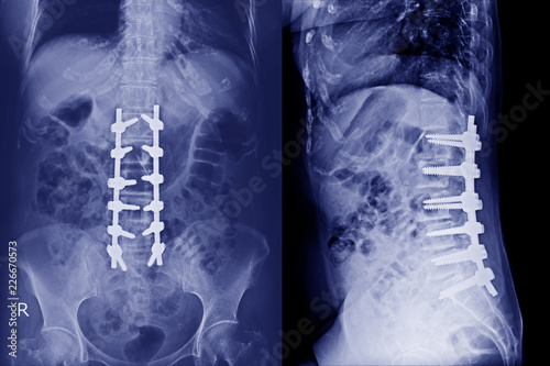
X-ray Lumbar spine AP,Lateral : A human and postoperative treatment for degenerative lumbar disc disease by decompression and fix by iron rod and screws.Blue tone. - Buy this stock photo and explore

Thoracic spine (AP view) | Radiology Reference Article | Radiopaedia.org

Thoracolumbar spine x-rays

AP or PA Projection in Lumbar Spine X-ray - RadTechOnDuty

GW700H956 700×956 pixels | Radiology student, Radiology, Radiology technologist

X-ray of dorsal spine: ap/lateral views showing diffuse soft-tissue... | Download Scientific Diagram
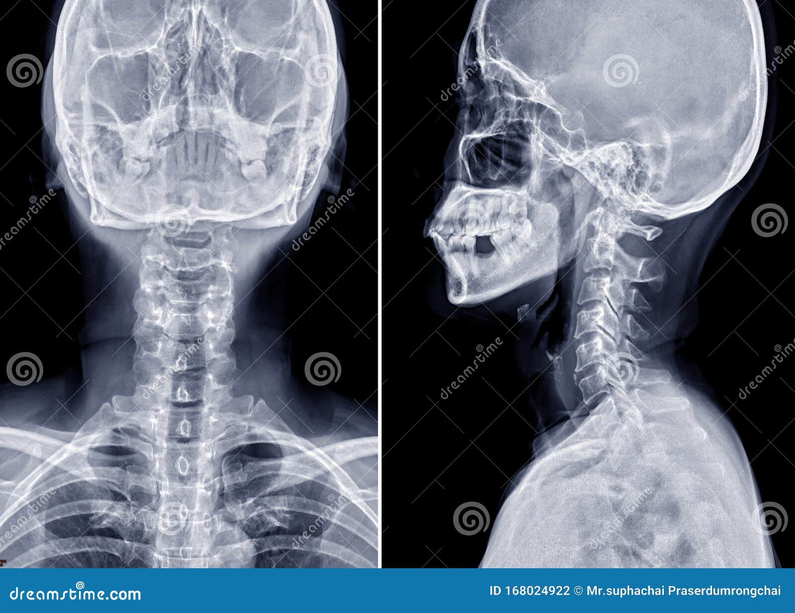
X-ray C-spine Or X-ray Image Of Cervical Spine AP And Lateral View Stock Photo - Image of cranium, illness: 168024922
Spine (Chapter 6) - Postgraduate Orthopaedics
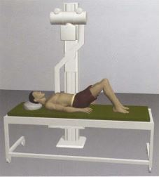
CE4RT - Radiographic Positioning of the Lumbar Spine for X-ray Techs

LATERAL POSITION: LUMBAR SPINE - RadTechOnDuty

Film X-ray Lumbar Spine , L-S Spine AP,Lateral View Grade Spondylolithesis.. Stock Photo, Picture And Royalty Free Image. Image 92218046.

Preoperative X-rays of dorsolumbar spine AP and lateral | Open-i

View Image
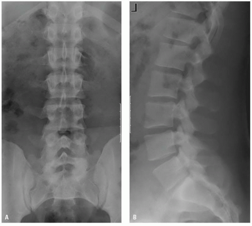
Imaging Thoracolumbar Spine Trauma | Radiology Key

X-Ray Positioning for Thoracic Spine - YouTube

Game Statistics - Lumbosacral Spine (AP and LAT)

AP Lumbar Spine X-Ray Diagram | Quizlet

Oblique Lumbar Spine X-ray Labeled (Page 1) - Line.17QQ.com
Shokeen X-ray & Dignostics Centre

Anteroposterior (AP; a) and lateral (b) radiographs of the lumbar spine... | Download Scientific Diagram
Posting Komentar untuk "xr lumbar spine ap and lateral"